Virtuel mikroskopi i oral patologi på tandlægestudiet
Den nye teknologi digital patologi giver øget fleksibilitet for både studerende og undervisere i patologiundervisningen, konkluderer forskere i denne fokusartikel.
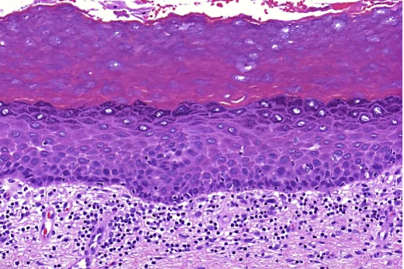
Undervisningen i oral patologi på tandlægestudiet har som mål, at de studerende skal få indsigt i histopatologien ved de mest normale sygdomme og tilstande, som de vil møde i deres senere karriere. En sådan kundskab er vigtig for deres forståelse af sygdommenes kliniske manifestationer og deres udvikling. Tidligere er kurset i oral patologi foregået på kursussale udstyret med mikroskop, hvilket har været ressourcekrævende og har givet de studerende begrænset tid til at studere vævssnittene. En ny teknologi er udviklet, hvor vævssnittene scannes og uploades til digitale platforme, der kan gøres tilgængelige for de studerende, som dermed kan studere vævssnittene i et virtuelt mikroskop. Dette giver øget fleksibilitet for både studerende og undervisere. De studerende kan forberede sig til undervisningen og kan senere studere vævssnittene på egen computer. Integrationen med de kliniske fag gøres enklere, og undervisningsmateriale kan deles mellem institutioner. De scannede vævssnit har en kvalitet, der svarer til et lysmikroskop af høj kvalitet.
Klinisk relevans:
Oral patologi er tæt knyttet til kliniske fag, særlig oral kirurgi og oral medicin. Det er vigtigt, at tandlægestuderende får viden om en forandrings histopatologiske udseende og forstår, hvordan dette reflekteres i det kliniske billede og udviklingen af en sygdom. Digital patologi giver nye muligheder for at integrere patologi i klinikken. Et eksempel kan være en patient, som kommer til kontrol, efter at man har taget en biopsi. Man kan da uploade det histopatologiske billede af biopsien og sammenholde det med det kliniske billede og således give de studerende en større forståelse for sammenhængen mellem vævets reaktionsmønster og de forskellige behandlingsmetoder og indgreb. Brugen af digital patologi i undervisningen vil skabe forudsætninger for, at fremtidens tandlæger får en større forståelse for oral patologi og dermed kan yde en bedre behandling. Forudsætningen er muligheder for at få scannet vævssnittene med høj kvalitet, og at de studerende har adgang til et sikkert digitalt system, hvor de scannede vævssnit er tilgængelige.Virtual microscopy in oral pathology at dental school
Teaching in oral pathology at the dental schools aims to give students insight into the histopathology of the most common diseases and conditions that they will meet later in their career. Such knowledge is important for their understanding of the clinical manifestation of the diseases and their development. Previously, courses in oral pathology have been arranged in special halls, equipped with microscopes, which has been resource-intensive and has given the students limited time to study the tissue sections. New technology has been developed where tissue sections are scanned and uploaded to digital platforms that can be made available to students, who may study the tissue sections as in a virtual microscope, leading to increased flexibility both for students and teachers. Students can prepare for the lectures and can later study the tissue sections on their own computer. The integration with clinical subjects is made easier, and teaching material can be shared between institutions. The scanned tissue sections have a quality compatible with a high quality light microscope.